Muller Cells - Muller Glia Are A Major Cellular Source Of Survival Signals For Retinal Neurons In Diabetes Diabetes - Muller glia, astroglia or astrocytes, and.
byAdmin•
0
Muller Cells - Muller Glia Are A Major Cellular Source Of Survival Signals For Retinal Neurons In Diabetes Diabetes - Muller glia, astroglia or astrocytes, and.. An alternate pathway of chromophore recycling which uses muller's cells within the retina. A single progenitor cell gives rise to both muller cells and retinal neurons(turner and cepko, 1987) although apparently in two phases. Classic birthdating studies showed that each of the major classes of retinal cells was generated at specific times during development, and in a specific order. Possible functions of muller cells during retinal development. We conclude that p27kip1 regulates müller glial cell proliferation during reactive gliosis.
We conclude that p27kip1 regulates müller glial cell proliferation during reactive gliosis. Muller cells express the neuronal progenitor cell marker nestin in both differentiated and undifferentiated human foetal retina. A few will be available to trace for triple points in eyewire for two days only: Müller glia, or müller cells, are glial cells found in the retina, which serve as support cells for the neurons of the retina as all glial cells do. An alternate pathway of chromophore recycling which uses muller's cells within the retina.
Frontiers Mirnas And Muller Glia Reprogramming During Retina Regeneration Cell And Developmental Biology from www.frontiersin.org Muller cell and neuronal remodeling in retinal detachment and reattachment and their potential consequences for visual recovery: Muller glia — müller glia, or müller cells, are glial cells found in the vertebrate retina, which normally serve the functions of cell — the basic structural and functional unit in people and all living things. Muller cells were first described in 1851 by heinrich muller 534. They are found in the vertebrate retina, which serve as support cells for the neurons, as all glial cells do. Müller cells, the major type of glial cells in the retina, are responsible for the homeostatic and metabolic support of retinal neurons. Pdf | müller glial cells span the entire thickness of the tissue, and ensheath all retinal neurons, in vertebrate retinae of all species. Cells, muller glia, muller cells (en); A few will be available to trace for triple points in eyewire for two days only:
The muller cell is the principal glial cell of the vertebrate retina;
Classic birthdating studies showed that each of the major classes of retinal cells was generated at specific times during development, and in a specific order. They are found in the vertebrate retina, which serve as support cells for the neurons, as all glial cells do. Muller cells span the entire width. The role of retinal muller cells in retinal function and retinal damage, a lecture given by prof. Muller cell and neuronal remodeling in retinal detachment and reattachment and their potential consequences for visual recovery: Keywords amacrine cell bipolar cell cone fovea horizontal cell muller cells photoreceptor retina retinal ganglion cell rod. Possible functions of muller cells during retinal development. In mammalian retina, three types of glia have been identified: A few will be available to trace for triple points in eyewire for two days only: A single progenitor cell gives rise to both muller cells and retinal neurons(turner and cepko, 1987) although apparently in two phases. Some articles on muller, cells, muller cell, cell, muller cells: Muller cells are glia found in the eyes of all mammals. We conclude that p27kip1 regulates müller glial cell proliferation during reactive gliosis.
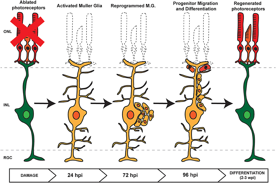
A single progenitor cell gives rise to both muller cells and retinal neurons(turner and cepko, 1987) although apparently in two phases. They are found in the vertebrate retina, which serve as support cells for the neurons, as all glial cells do. They are the most common type of glial cell found in the retina. Cells, the muller cell (fig. However, following injury to the retina, it has been shown in.
Muller Cells In The Healthy Retina Springerlink from media.springernature.com A review and reconsideration of recent data. Pdf | müller glial cells span the entire thickness of the tissue, and ensheath all retinal neurons, in vertebrate retinae of all species. Each cell is a small. Classic birthdating studies showed that each of the major classes of retinal cells was generated at specific times during development, and in a specific order. A single progenitor cell gives rise to both muller cells and retinal neurons(turner and cepko, 1987) although apparently in two phases. Muller glia — müller glia, or müller cells, are glial cells found in the vertebrate retina, which normally cell — the basic structural and functional unit in people and all living things. Muller cell and neuronal remodeling in retinal detachment and reattachment and their potential consequences for visual recovery: Müller cells, the major type of glial cells in the retina, are responsible for the homeostatic and metabolic support of retinal neurons.
Muller glia — müller glia, or müller cells, are glial cells found in the vertebrate retina, which normally serve the functions of cell — the basic structural and functional unit in people and all living things.
Retinal glial cells are usually subdivided into macroglia (muller cells and astrocytes) and microglia with specific morphological, physiological, and antigenic characteristics. Müller cells, the major type of glial cells in the retina, are responsible for the homeostatic and metabolic support of retinal neurons. Ido perlman of technion during the conference from basic. Muller cells span the entire width. An alternate pathway of chromophore recycling which uses muller's cells within the retina. By mediating transcellular ion, water, and bicarbonate transport, müller. Müller glial cells are the major support cell for neurons in the vertebrate retina. A review and reconsideration of recent data. The role of retinal muller cells in retinal function and retinal damage, a lecture given by prof. Possible functions of muller cells during retinal development. Muller glia — müller glia, or müller cells, are glial cells found in the vertebrate retina, which normally cell — the basic structural and functional unit in people and all living things. Muller glia, astroglia or astrocytes, and. A single progenitor cell gives rise to both muller cells and retinal neurons(turner and cepko, 1987) although apparently in two phases.
Keywords amacrine cell bipolar cell cone fovea horizontal cell muller cells photoreceptor retina retinal ganglion cell rod. Müller glial cells are the major support cell for neurons in the vertebrate retina. In mammalian retina, three types of glia have been identified: A few will be available to trace for triple points in eyewire for two days only: Muller glia — müller glia, or müller cells, are glial cells found in the vertebrate retina, which normally serve the functions of cell — the basic structural and functional unit in people and all living things.
Muller Glia And Phagocytosis Of Cell Debris In Retinal Tissue Bejarano Escobar 2017 Journal Of Anatomy Wiley Online Library from onlinelibrary.wiley.com By mediating transcellular ion, water, and bicarbonate transport, müller. A review and reconsideration of recent data. Müller cells, the major type of glial cells in the retina, are responsible for the homeostatic and metabolic support of retinal neurons. Pdf | müller glial cells span the entire thickness of the tissue, and ensheath all retinal neurons, in vertebrate retinae of all species. They are found in the vertebrate retina, which serve as support cells for the neurons, as all glial cells do. Muller glia, astroglia or astrocytes, and. The muller cell is the principal glial cell of the vertebrate retina; We conclude that p27kip1 regulates müller glial cell proliferation during reactive gliosis.
Ido perlman of technion during the conference from basic.
Pdf | müller glial cells span the entire thickness of the tissue, and ensheath all retinal neurons, in vertebrate retinae of all species. In mammalian retina, three types of glia have been identified: A review and reconsideration of recent data. Cells, muller glia, muller cells (en); Müller glia, or müller cells, are a type of retinal glial cells, first recognized and described by heinrich müller. Müller cells, the principal glial cells of the retina, play an important role in immune responses. They are found in the vertebrate retina, which serve as support cells for the neurons, as all glial cells do. These elongated cells run from the outer limiting membrane to the inner limiting membrane. Ido perlman of technion during the conference from basic. The following 7 files are in this category, out of 7 total. A single progenitor cell gives rise to both muller cells and retinal neurons(turner and cepko, 1987) although apparently in two phases. Cells, the muller cell (fig. We conclude that p27kip1 regulates müller glial cell proliferation during reactive gliosis.
Muller cell and neuronal remodeling in retinal detachment and reattachment and their potential consequences for visual recovery: muller. Possible functions of muller cells during retinal development.