Renal Blood Vessels Labeled / Cyprinus sp. Kidney. Transverse section. 250X - Cyprinus ... / These vessels transport blood cells, nutrients, and oxygen to the tissues of the body.
byAdmin•
0
Renal Blood Vessels Labeled / Cyprinus sp. Kidney. Transverse section. 250X - Cyprinus ... / These vessels transport blood cells, nutrients, and oxygen to the tissues of the body.. Exposure of blood vessel organoids to hyperglycaemia and inflammatory cytokines in vitro induces thickening of the vascular basement membrane. These vessels transport blood cells, nutrients, and oxygen to the tissues of the body. The renal arteries arise, one on each side, from the abdominal aorta at a point opposite the upper border of the second lumbar this structure, called the renal corpuscular capsule , or bowman's capsule, encloses a cluster of capillaries (microscopic blood vessels) called. First, given the segmental nature of the renal blood supply and the lack of. It may require some insight to orient yourself on this 7.
This artery branches into the segmental arteries then the interlobar arteries, arcuate that depends on which what kind of blood vessel you cut, and how much of it is damaged. Put simply, they are supplied and drained by the branches of three primary vessels: The arteries are obscured by the renal veins in this image; The process of tubular secretion helps to secrete the urea from the blood to the collecting duct which is finally excreted in form of urine. Automatic blood vessel segmentation in the images can help speed diagnosis and improve the diagnostic performance of less specialized physicians.
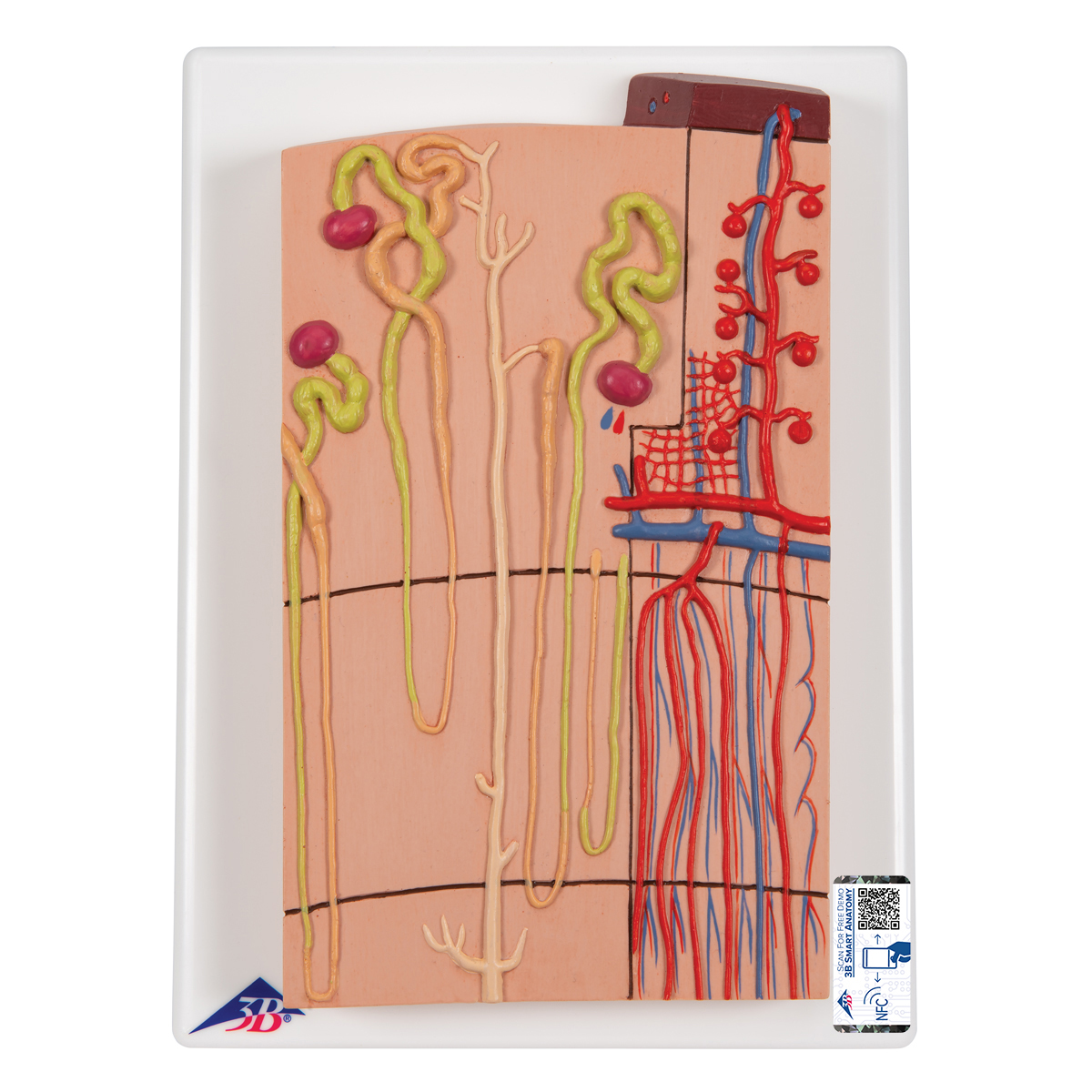
Anatomical Teaching Model - Plastic Anatomy Models - Renal ... from www.a3bs.com These give off a series of branches which enter the cortex as interlobular arterioles. (see also overview of blood vessel disorders of the kidneys.) there are two renal arteries—one supplies blood to the right kidney, the other to the left kidney. Note their relationship with the renal pelvis and ureters. These vessels transport blood cells, nutrients, and oxygen to the tissues of the body. Blood vessels (note outlines of red blood cells in slide 204) are also seen. Ready to learn about the blood vessels of the abdomen and pelvis (the abdominopelvic blood vessels)? Automatic blood vessel segmentation in the images can help speed diagnosis and improve the diagnostic performance of less specialized physicians. Exposure of blood vessel organoids to hyperglycaemia and inflammatory cytokines in vitro induces thickening of the vascular basement membrane.
Observe the distribution of blood vessels.
Renal vessels arise at the level of the intervertebral disc between l1 and l2 vertebrae. The innervation of the renal blood vessels. (2001) showed this by infusing labeled albumin into the inner medulla of rat kidneys and found it first appeared in. The arteries are mostly posterior to the veins. You can support the work of campbellteaching, at no cost whatsoever to yourself, if you use the link below as your bookmark to access amazon. Example, the venous blood passes through interlobular, arcuate, interlobar, and renal veins. The process of tubular secretion helps to secrete the urea from the blood to the collecting duct which is finally excreted in form of urine. The renal cortex and medulla contain a complex network of blood vessels. They also take waste and carbon dioxide away from the tissues. (see also overview of blood vessel disorders of the kidneys.) there are two renal arteries—one supplies blood to the right kidney, the other to the left kidney. Blood vessels labeled simple : Blood vessels associated with the kidneys and adrenal glands. This is an online quiz called renal blood vessels.
You can support the work of campbellteaching, at no cost whatsoever to yourself, if you use the link below as your bookmark to access amazon. August 17, 2020 so, you want to learn. Morphology of renal lymph vessels. Blood vessels (note outlines of red blood cells in slide 204) are also seen. Bloodvessel — the blood vessels are part of the circulatory system and function to transport blood throughout the body.
Photomicrograph of the kidney showing were renal blood ... from www.researchgate.net Morphology of renal lymph vessels. Blood enters the renal vascular system through the renal artery. Edged by blood vessels running between lobes is an arter and vein. This artery branches into the segmental arteries then the interlobar arteries, arcuate that depends on which what kind of blood vessel you cut, and how much of it is damaged. Put simply, they are supplied and drained by the branches of three primary vessels: First, given the segmental nature of the renal blood supply and the lack of. • identification of blood vessels as arteries, capillaries or veins from the structure of their walls. Study renal blood flow and gfr flashcards from hanifa ahmed's university of leicester class online, or in brainscape's iphone or android app.
Observe the distribution of blood vessels.
The same applies to the thoracic aorta that runs through the chest and other arteries such as the renal artery or pelvic artery which supply the kidneys and pelvic area. The most important vessels in the system are the capillaries , the microscopic vessels which enable the actual exchange of water and … … The process of tubular secretion helps to secrete the urea from the blood to the collecting duct which is finally excreted in form of urine. The renal vein then joins the inferior vena cava as it courses through the abdominal cavity. ✓ learn faster with spaced repetition. Development and function of the blood vessels: Exposure of blood vessel organoids to hyperglycaemia and inflammatory cytokines in vitro induces thickening of the vascular basement membrane. Observe the distribution of blood vessels. It carries the urea loaded blood into the glomerulus of the kidney. These arteries branch into many smaller arteries. The renal arteries arise, one on each side, from the abdominal aorta at a point opposite the upper border of the second lumbar this structure, called the renal corpuscular capsule , or bowman's capsule, encloses a cluster of capillaries (microscopic blood vessels) called. The blood vessels make up the body's cardiovascular system. The interlobar arteries which pass between the renal pyramids, arch around the base of the pyramid as the arcuate arteries.
A gradual narrowing of one or both of the renal arteries may cause high blood pressure or a. The arteries are mostly posterior to the veins. The best websites voted by users. Blood vessels (note outlines of red blood cells in slide 204) are also seen. ✓ learn faster with spaced repetition.
Annotated diagrams showing the nephrons up close from mammothmemory.net These vessels transport blood cells, nutrients, and oxygen to the tissues of the body. ✓ learn faster with spaced repetition. Dimitrios mytilinaios md, phd • last reviewed: This is an online quiz called renal blood vessels. Morphology of renal lymph vessels. The blood supply to the kidneys originates from the paired renal arteries, which branch into segmental arteriesat the renal hilum. The renal cortex and medulla contain a complex network of blood vessels. This artery branches into the segmental arteries then the interlobar arteries, arcuate that depends on which what kind of blood vessel you cut, and how much of it is damaged.
Blood enters the renal vascular system through the renal artery.
The renal vein then joins the inferior vena cava as it courses through the abdominal cavity. Observe the distribution of blood vessels. The renal arteries arise, one on each side, from the abdominal aorta at a point opposite the upper border of the second lumbar this structure, called the renal corpuscular capsule , or bowman's capsule, encloses a cluster of capillaries (microscopic blood vessels) called. Which of the labeled ultrastructural features most significantly impedes the passage of negatively charged molecules? Choose from 500 different sets of flashcards about renal blood vessels on quizlet. Ready to learn about the blood vessels of the abdomen and pelvis (the abdominopelvic blood vessels)? The renal cortex and medulla contain a complex network of blood vessels. A gradual narrowing of one or both of the renal arteries may cause high blood pressure or a. Exposure of blood vessel organoids to hyperglycaemia and inflammatory cytokines in vitro induces thickening of the vascular basement membrane. (see also overview of blood vessel disorders of the kidneys.) there are two renal arteries—one supplies blood to the right kidney, the other to the left kidney. You can support the work of campbellteaching, at no cost whatsoever to yourself, if you use the link below as your bookmark to access amazon. We'll assume for the purposes of this answer that the. The most important vessels in the system are the capillaries , the microscopic vessels which enable the actual exchange of water and … …
Exposure of blood vessel organoids to hyperglycaemia and inflammatory cytokines in vitro induces thickening of the vascular basement membrane blood vessels labeled. Blood vessels (note outlines of red blood cells in slide 204) are also seen.


.jpg)