Human Leg Bone Diagram : Free Vector Human Knee Anatomy Diagram / Human anatomy for muscle, reproductive, and skeleton.
byAdmin•
0
Human Leg Bone Diagram : Free Vector Human Knee Anatomy Diagram / Human anatomy for muscle, reproductive, and skeleton.. When your muscles contract, they pull the bone they're. The human leg, in the general word sense, is the entire lower limb of the human body, including the foot, thigh and even the hip or gluteal region. The hip joint is the uppermost part of the leg where the head of the thigh bone (femur) fits into the socket of the pelvis. Legs come in all shapes and sizes, ranging from portly and stout, to the streamlined, almost artists usually begin their study of the legs by focusing on the athletic type, because the shapes of the muscles are more easily seen. The foot bones shown in this diagram are the talus, navicular, cuneiform, cuboid, metatarsals and calcaneus.
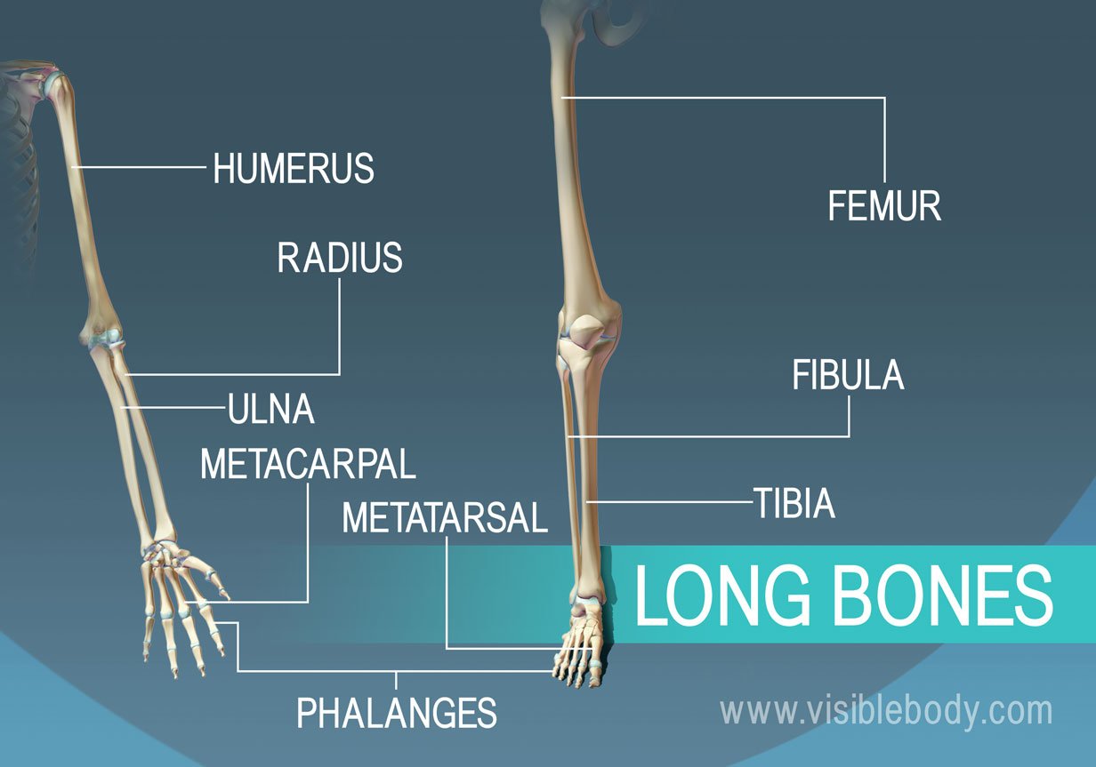
Includes leg (femur, tibia, patella, and fibula) and foot (tarsals and digits) bones. This includes the foot, thigh and even the hip or gluteal region. Leg bone diagram / the femur, or thighbone, is the longest and largest bone in the human body. In an erect posture, you can place the pelvic bone (a narrower version of the head's egg) this completes the basic, undifferentiated human proportions, and here's a diagram to sum up all of. Includes obj for maximum compatibility.
Types Of Bones Learn Skeleton Anatomy from www.visiblebody.com High resolution textures and displacement included. How to draw human skull front/profile | human anatomy. Distal end of right humerus. The knee joint is the largest joint in the body and is primarily a hinge joint. Find stockbilleder af infographic diagram human femur bone leg i hd og millionvis af andre royaltyfri stockbilleder, illustrationer og vektorer i shutterstocks samling. The human leg consists of 8 bones, 4 per leg. Human bones diagram (page 1). Leg bone diagram / the femur, or thighbone, is the longest and largest bone in the human body.
In an erect posture, you can place the pelvic bone (a narrower version of the head's egg) this completes the basic, undifferentiated human proportions, and here's a diagram to sum up all of.
Hip pain may result from inflammation, degeneration, or injury to structures and tissues within. Human leg bones 3d model. Bones in the hand and wrist right hand. These bones are arranged into two major divisions: The knee joint is the largest joint in the body and is primarily a hinge joint, although. Its decrease finish helps create the knee joint. High quality realistic skeleton legs. Joints of hand anterior view, lateral view, right hand. The human leg, in the general word sense, is the entire lower limb of the human body, including the foot, thigh and even the hip or gluteal region. Related posts of diagram of leg bones. The knee joint is the largest joint in the body and is primarily a hinge joint. Rated 5.00/5 based on 2 votes. This diagram depicts human leg bone anatomy.
There also are bands of fibrous connective tissue—the ligaments and the tendons—in intimate relationship with the parts of the skeleton. The first scientist to correctly illustrate the human skeleton with all of its bones. Human anatomy for muscle, reproductive, and skeleton. Bones in the hand and wrist right hand. Separate files for each identical bones.
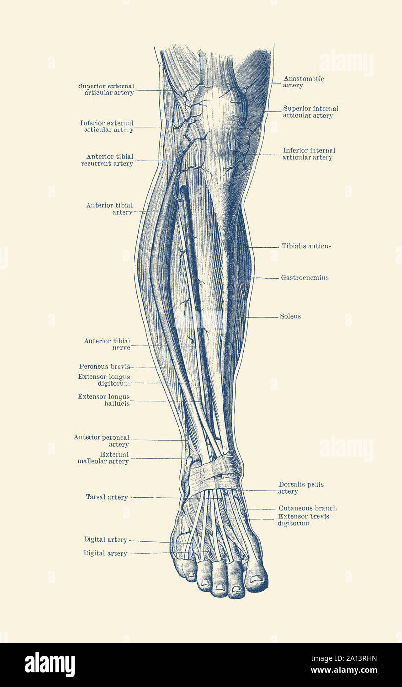
Vintage Anatomy Print Of The Human Leg Showcasing The Veins And Arteries Stock Photo Alamy from c8.alamy.com Human anatomy for muscle, reproductive, and skeleton. Separate files for each identical bones. The human leg, in the general word sense, is the entire lower limb of the human body, including the foot, thigh and even the hip or gluteal region. Distal end of right humerus. The foot bones shown in this diagram are the talus, navicular, cuneiform, cuboid, metatarsals and calcaneus. Learn vocabulary, terms and more with flashcards, games and other study tools. This framework consists of many individual bones and cartilages. It joins with the scapula above at the shoulder joint (or.
The first scientist to correctly illustrate the human skeleton with all of its bones.
This framework consists of many individual bones and cartilages. However, the definition of human anatomy mentions only to the section of the lower limb extending from the knee to the ankle, also known as the crus. Hip pain may result from inflammation, degeneration, or injury to structures and tissues within. Disposition of rotator cuff muscles diagram. The bones of your leg have roughened patches on their surfaces where muscles are attached. File is ready to render. They are named by region formed by the left and right hip bones, the pelvic girdle connects the lower limb (leg) bones to the axial skeleton. The human leg consists of 8 bones, 4 per leg. The hip joint is the uppermost part of the leg where the head of the thigh bone (femur) fits into the socket of the pelvis. Human anatomy for muscle, reproductive, and skeleton. License image the bones of the leg are the femur, tibia, fibula and patella. The foot bones shown in this diagram are the talus, navicular, cuneiform, cuboid, metatarsals and calcaneus. The human leg consists of 8 bones, 4 per leg.
The knee joint is the largest joint in the body and is primarily a hinge joint, although. This diagram depicts human leg bone anatomy. Find stockbilleder af infographic diagram human femur bone leg i hd og millionvis af andre royaltyfri stockbilleder, illustrationer og vektorer i shutterstocks samling. How to draw human skull front/profile | human anatomy. When you stand or walk, all the weight of your upper body rests on them.
Free Vector Human Knee Anatomy Diagram from image.freepik.com Separate files for each identical bones. These bones are arranged into two major divisions: There also are bands of fibrous connective tissue—the ligaments and the tendons—in intimate relationship with the parts of the skeleton. Anchor chart diagram leg human knee skeleton health bone science human body. Foot and ankle diagram anatomy. They are named by region formed by the left and right hip bones, the pelvic girdle connects the lower limb (leg) bones to the axial skeleton. Bones in the hand and wrist right hand. How to draw human skull front/profile | human anatomy.
Human bones diagram (page 1).
Related posts of diagram of leg bones. The femur, or thighbone, is the longest and largest bone within the human physique. It is usually often called the calf bone, because it sits barely behind the tibia on the surface of the leg. They are named by region formed by the left and right hip bones, the pelvic girdle connects the lower limb (leg) bones to the axial skeleton. Disposition of rotator cuff muscles diagram. He leg's main function in the human is for locomotion and support of use the leg bones diagrams to learn the names of the leg bones and leg anatomy. 3d model of human leg bones available for download in fbx, obj, 3ds, c4d and other file formats for 23 software. When your muscles contract, they pull the bone they're. Learn vocabulary, terms and more with flashcards, games and other study tools. High quality realistic skeleton legs. Upper leg bones diagram the junction of where these structures converge at the pubic bone. Bones in the hand and wrist right hand. Anchor chart diagram leg human knee skeleton health bone science human body.
Human bones diagram (page 1) leg bone diagram. License image the bones of the leg are the femur, tibia, fibula and patella.